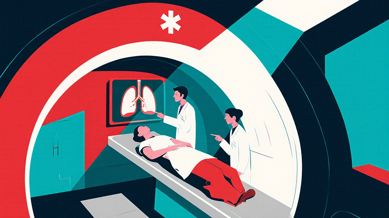
Contrast CT: What It Is and Why It Matters
When you hear contrast CT, a scan that combines a traditional computed tomography (CT) exam with an injected iodine‑based contrast agent to highlight blood vessels, organs, and lesions. Also known as contrast‑enhanced CT, it provides doctors with clearer images for diagnosing cancers, vascular disease, and trauma, the technique can be a game‑changer in modern medicine. contrast CT is not just a fancy term; it’s a practical tool that turns ordinary slices into diagnostic gold mines.
Key Players Behind a Successful Scan
The magic starts with a contrast agent, an iodine‑rich fluid injected into the bloodstream to make vessels and tissues stand out on the images. Without it, many subtle abnormalities remain hidden. The next piece is computed tomography, the high‑speed X‑ray technology that captures cross‑sectional images in seconds. Together, they create a detailed 3‑D map that radiologists interpret. In the broader picture, radiology, the medical specialty focused on imaging diagnostics, relies on these tools to guide treatment plans. And because the end goal is to give clinicians actionable information, diagnostic imaging, the umbrella term for all visual tests, frames the whole process.
Understanding how these pieces fit together clears up common confusion. For instance, many wonder whether the contrast agent poses risks; the answer hinges on patient history and kidney function, not on the technology itself. Likewise, the CT scanner’s speed and resolution determine how well the agent’s benefits are captured, so a modern machine often means sharper images with lower radiation doses. In practice, a radiology team evaluates the need for contrast based on the clinical question – a lung nodule, a suspected aneurysm, or a hidden tumor. This decision‑making loop—patient assessment, contrast selection, CT acquisition, radiology interpretation—creates a seamless workflow that improves diagnostic confidence.
All of this matters because the articles you’ll find below dive deep into real‑world scenarios where contrast CT shines. From exploring how specific medications like Atazanavir affect pulmonary health to comparing treatment options for diseases that show up on scans, the collection gives you a practical roadmap. Whether you’re a clinician looking for monitoring tips, a patient curious about what to expect during the exam, or just someone wanting to understand the technology, the posts cover safety guidelines, cost considerations, and the latest research findings. Let’s move on to the detailed guides that will help you make sense of contrast‑enhanced imaging in everyday practice.
CT Scans in Embolism Diagnosis and Management: What You Need to Know
Explore how CT scans detect and guide treatment of embolisms, from pulmonary clots to arterial blockages, with practical tips, pitfalls, and a CTA vs V/Q comparison.
Read more