Contrast Dose Calculator
Contrast Dose Calculator
Calculate the appropriate iodinated contrast dose based on patient weight. This is essential for safe and effective CT angiography.
Enter patient weight to calculate appropriate contrast dose.
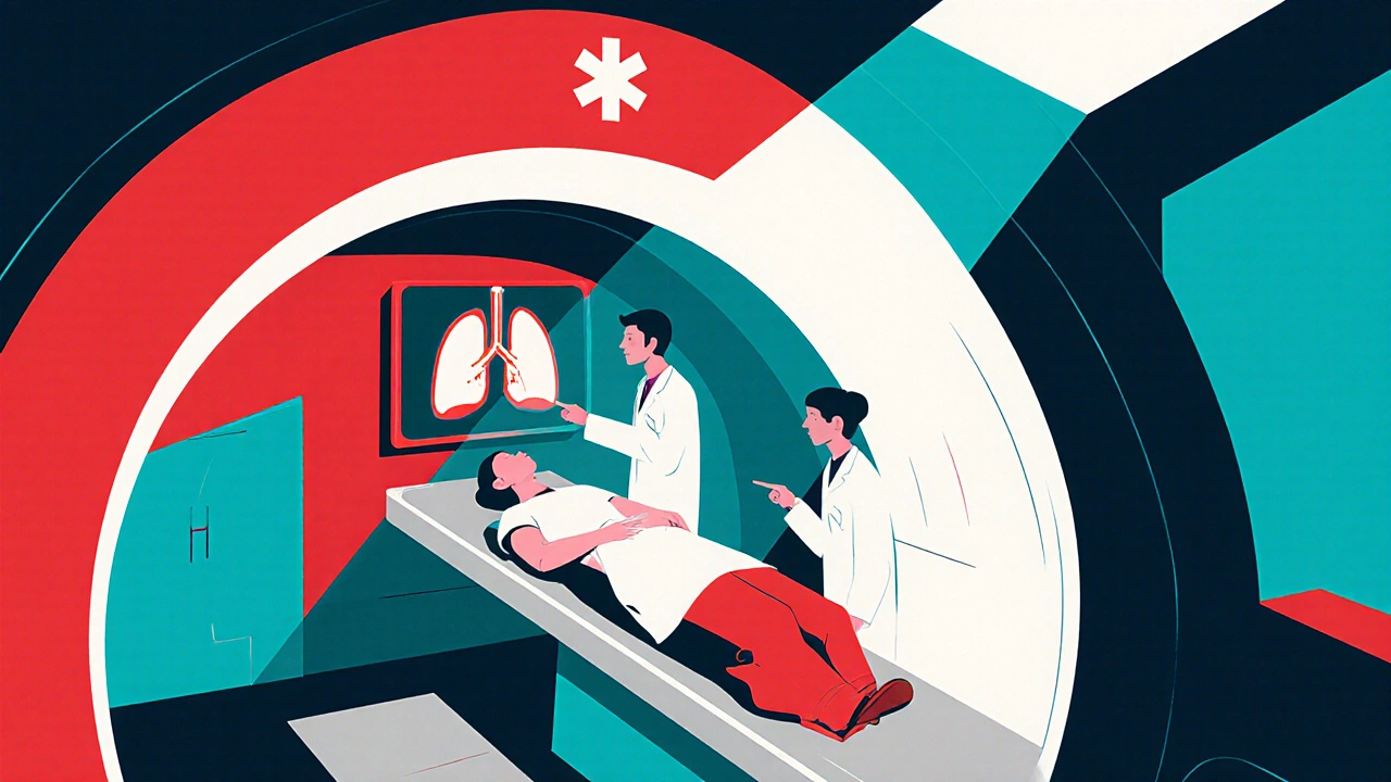
When a clot blocks a blood vessel, every second counts. Doctors need a fast, reliable way to see where the blockage is and decide how to treat it. That’s where CT scans step in. In the next few minutes you’ll learn how the technology works, what kinds of embolism it can spot, and how the images guide life‑saving decisions.
How a CT Scan Generates Images
CT scan is a cross‑sectional X‑ray technique that creates detailed 3‑D pictures of the inside of the body. A rotating X‑ray tube fires thin beams through the patient while detectors capture the attenuation rates. A computer stitches the data into slices that can be viewed individually or combined into a volumetric view.
The key metric is the Hounsfield unit (HU), which quantifies how much the X‑ray beam is weakened by different tissues. Water is set at 0 HU, air at -1000 HU, and dense bone up to +1000 HU. By measuring HU, radiologists can differentiate blood, clot, and surrounding organs.
Why CT Is the Go‑to Modality for Embolism
Emboli are often small, mobile, and located in critical vessels. CT provides three advantages that make it a frontline tool:
- Speed - Modern scanners finish a chest CT in under a minute, which is crucial in emergencies.
- Resolution - Sub‑millimeter detail lets radiologists spot clots as thin as a few millimetres.
- Contrast capability - Intravenous iodinated contrast highlights the blood pool, making a blockage stand out as a filling defect.
Because the scan can be performed in the emergency department, it fits neatly into the acute care workflow.
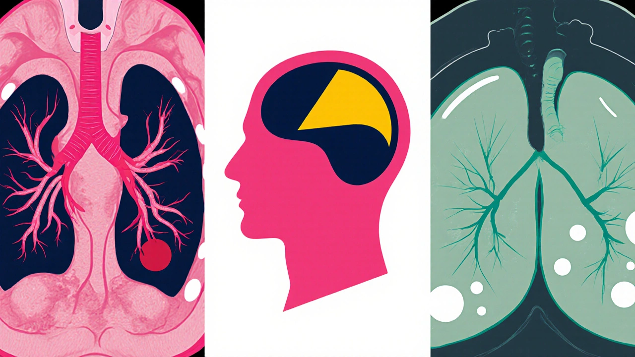
Types of Embolism Detectable by CT
Not all emboli look the same. Here’s a quick rundown of the most common ones that CT can reveal.
- Pulmonary embolism is a clot that travels to the arteries of the lungs, often originating from a deep vein thrombosis (DVT).
- Arterial embolism blocks blood flow to an organ, such as a brain (causing stroke) or kidney.
- Fat embolism appears after long‑bone fractures; CT shows a characteristic “ground‑glass” pattern in the lungs.
- Air embolism occurs when air enters the circulation, typically seen as low‑density gas bubbles on CT.
Each type has a distinct appearance, but all rely on contrast‑enhanced CT to confirm the diagnosis.
Preparing for a Contrast‑Enhanced CT Scan
Before the scanner fires, the patient receives an iodinated contrast agent through an IV. The timing of the injection determines which vascular phase is captured:
- Arterial phase - Captured 20‑30 seconds after injection; ideal for detecting arterial emboli.
- Pulmonary artery phase - 60‑70 seconds post‑injection; optimizes pulmonary embolism visualization.
- Venous phase - 2‑3 minutes later; useful for assessing deep vein thrombosis in the pelvis.
Patients with kidney impairment may need a reduced‑dose protocol or an alternative imaging modality such as magnetic resonance angiography (MRA).
Interpreting CT Findings: What Radiologists Look For
After the scan, the radiologist reviews the images on a high‑resolution workstation. Key signs include:
- Filling defect - A dark wedge‑shaped area within a bright, contrast‑filled vessel suggests a clot.
- V/Q mismatch - Areas of lung that receive blood but not ventilation appear as peripheral opacities.
- Right‑heart strain - Enlarged right ventricle or a bowing interventricular septum indicates pressure overload from a large PE.
Radiologists also measure the clot’s length and location, which directly influences treatment choices.
Management Pathway After CT Confirms an Embolism
Once the CT report is in hand, the clinical team moves quickly to treat the clot. The main options are:
- Anticoagulation - Heparin, low‑molecular‑weight heparin (LMWH), or direct oral anticoagulants (DOACs) prevent further clot growth.
- Thrombolysis - In massive pulmonary embolism with hemodynamic collapse, tissue‑type plasminogen activator (tPA) can dissolve the clot within hours.
- Catheter‑directed therapy - For sub‑massive PE, a catheter can mechanically fragment or aspirate the clot.
The CT also helps risk‑stratify patients. A small, peripheral PE may be managed with oral anticoagulants alone, while a central saddle embolus often triggers more aggressive intervention.
CT Angiography vs. Ventilation‑Perfusion Scan: A Quick Comparison
| Aspect | CT Angiography | Ventilation‑Perfusion Scan |
|---|---|---|
| Speed | 1-2 minutes (single‑breath hold) | 15-20 minutes (multiple breaths) |
| Radiation dose | ≈4-7 mSv (lower with modern scanners) | ≈2-3 mSv |
| Contrast needed | Yes - iodinated contrast | No |
| Accuracy | Sensitivity > 95 %, specificity ≈ 90 % | Sensitivity ≈ 80 %, specificity ≈ 70 % |
| Best for | Acute PE, detailed anatomy, surgical planning | Patients with contrast allergy or renal failure |
In most hospitals, CTA is the first‑line test because it delivers rapid, high‑resolution images. The V/Q scan remains valuable when contrast can’t be used.
Practical Checklist for Clinicians Ordering a CT for Embolism
- Confirm indication (suspected PE, stroke, limb ischemia).
- Review renal function; calculate eGFR.
- Ask about contrast allergies; have pre‑medication ready if needed.
- Choose the appropriate scan phase (arterial, pulmonary, venous).
- Ensure IV access of at least 18‑gauge for rapid contrast injection.
- Document patient weight for contrast dose calculation.
- Post‑scan, verify that the radiology report includes clot size, location, and any signs of right‑heart strain.
Following this list reduces repeat scans and speeds up the treatment decision.
Common Pitfalls and How to Avoid Them
Even with a powerful tool like CT, mistakes happen. Here are three frequent issues and quick fixes:
- Timing errors - If the scan is performed too early, contrast may not have reached the target vessels. Use automated bolus‑tracking software to trigger the scan at the right moment.
- Motion artifacts - Shallow breathing or coughing blurs the images. Coach the patient to hold a deep breath for the brief acquisition period.
- Misinterpreting beam‑hardening - Dense bone can create streaks that mimic filling defects. Adjust the window level or request a reconstruction with metal‑artifact reduction.
Future Directions: AI‑Assisted CT Interpretation
Artificial intelligence is already being piloted to flag potential emboli automatically. Early studies show AI can detect pulmonary embolism with sensitivity above 96 % and cut reading time by half. While the technology is not yet standard, clinicians should stay aware of emerging software that could become part of routine workflow.
Can a CT scan miss a small pulmonary embolism?
Yes, very tiny segmental clots can be overlooked, especially if the contrast timing is off or if there is significant motion artifact. However, modern multi‑detector CT scanners reduce this risk to less than 5 % when protocols are followed correctly.
What if I can’t receive iodinated contrast?
In cases of severe contrast allergy or renal insufficiency, a ventilation‑perfusión (V/Q) scan or magnetic resonance angiography (MRA) may be used. These alternatives avoid iodine but have their own limitations in resolution and availability.
How long does a CT‑based diagnosis influence treatment?
The CT report is most critical during the acute phase (first 6 hours). After that, the imaging helps guide long‑term anticoagulation duration and follow‑up imaging strategy.
Is there radiation risk for repeated CT scans?
Repeated exposure does add cumulative dose, but modern dose‑reduction techniques keep each chest CTA under 5 mSv, roughly equivalent to 1.5 years of natural background radiation. Clinicians weigh the benefit of accurate diagnosis against this modest risk.
What are the signs of right‑heart strain on CT?
Look for an enlarged right ventricle, a septal bowing toward the left ventricle, and reflux of contrast into the hepatic veins. These findings suggest a large central embolus and may prompt thrombolytic therapy.
Bret Toadabush
October 22, 2025 AT 18:18Those CT scans are just a way for the gov to sneak extra radiation into our bodies, no doubt.
Diane Thurman
October 27, 2025 AT 08:25I cant believe how many people just accept the "official" guidelines without questioning them. The article misses the point about the long‑term risks, and the data they quote is old. It's a bit of a lazy overview, honestly.
Christa Wilson
October 31, 2025 AT 23:32Great overview! 😃 This really helps demystify how CT angiography works and why it's so crucial in emergencies. Keep the info coming! 👍
Sameer Khan
November 5, 2025 AT 14:38The utilization of computed tomography in the assessment of embolic phenomena represents a paradigm shift in acute vascular medicine. Contemporary multidetector CT scanners achieve sub‑millimeter spatial resolution, thereby permitting visualization of intraluminal filling defects with unprecedented fidelity. Contrast timing, governed by bolus‑tracking algorithms, ensures optimal arterial phase acquisition, which is essential for discerning pulmonary arterial occlusions from adjacent parenchymal artifacts. Quantitative Hounsfield unit analysis further differentiates thrombus from surrounding blood, facilitating objective measurement of clot burden. In the context of pulmonary embolism, the presence of a central saddle embolus correlates with right‑ventricular strain indices, such as septal bowing and ventricular enlargement, as delineated on axial reconstructions. These morphologic markers possess prognostic significance and inform the decision to initiate systemic thrombolysis versus anticoagulation alone. Moreover, the integration of AI‑driven segmentation tools has been shown to reduce interpretation time by approximately 45 % while maintaining a sensitivity exceeding 96 %. Nonetheless, such algorithms require rigorous validation across diverse patient populations before routine deployment. The radiation dose associated with a chest CTA, currently mitigated through iterative reconstruction techniques, typically remains below 5 mSv, a value that must be weighed against the clinical urgency of definitive diagnosis. Renal insufficiency continues to pose a limitation; low‑dose contrast protocols or alternative modalities such as magnetic resonance angiography may be employed, albeit with trade‑offs in spatial resolution. From a workflow perspective, the rapid acquisition (<1 minute) enables seamless integration within emergency department pathways, minimizing delays to therapeutic intervention. The article's discussion of the CT versus V/Q paradigm is accurate, yet it understates the logistical constraints of scanner availability in resource‑limited settings. Interdisciplinary communication between radiology, emergency medicine, and vascular surgery remains paramount to optimize patient outcomes. Finally, ongoing advancements in photon‑counting detector technology promise further enhancements in contrast discrimination and dose reduction, heralding the next generation of embolism imaging.
Harini Prakash
November 10, 2025 AT 05:45I really appreciate the depth of your explanation; it makes the technical aspects feel more accessible. The note about AI tools is especially encouraging, as they could free up radiologists for more nuanced cases. 🌼
Rachael Turner
November 14, 2025 AT 20:52Interesting points you raised about the guidelines. I think it's worth noting that the risk‑benefit analysis varies with patient age and comorbidities. Also, the evolution of low‑dose protocols has changed the conversation.
Don Goodman-Wilson
November 19, 2025 AT 11:58Oh great, another glorified x‑ray that the pharma industry wants us to trust blindly.
Iris Joy
November 24, 2025 AT 03:05While the skepticism is understandable, the evidence supporting rapid CT in life‑threatening embolism is quite robust, and early detection can literally save lives.
John Connolly
November 28, 2025 AT 18:12CT angiography remains the workhorse for acute embolic evaluation; its speed and detail are unmatched, making it indispensable in modern emergency departments.
Emma Parker
December 3, 2025 AT 09:18yeah u r right the ct scans are super helpful but sometimes the wait time can be rly long lol
Joe Waldron
December 8, 2025 AT 00:25Indeed, the integration of high‑resolution CT imaging, contrast dynamics, and post‑processing algorithms, all contribute to a comprehensive diagnostic framework; however, clinicians must remain vigilant regarding potential pitfalls, such as motion artifacts, suboptimal bolus timing, and beam‑hardening effects, which can obscure critical findings.
Benedict Posadas
December 12, 2025 AT 15:32Great points! 😊 Just remember to double‑check the timing protocols – a tiny miss can hide a big clot! ;)
Kiara Gerardino
December 17, 2025 AT 06:38The moral imperative to employ every available tool in the fight against unseen killers cannot be overstated; to ignore the power of CT is to flirt with negligence.
Tim Blümel
December 21, 2025 AT 21:45Absolutely, the urgency of accurate imaging aligns with our ethical duty to patients; the data you presented underscores that responsibility. 💡
Emily Collins
December 26, 2025 AT 12:52While the philosophical considerations are compelling, the practical implementation hinges on protocol adherence and interdisciplinary communication.