Cancer Staging Calculator
Enter the details below to determine the cancer stage using the TNM system:
Quick Take
- tumor growth drives the transition from a harmless cell cluster to invasive cancer.
- Cancer is staged from I to IV using the TNM system (Tumor size, Node involvement, Metastasis).
- Metastasis marks the shift to stageIV and dramatically lowers survival odds.
- Key biological processes - angiogenesis, oncogene activation, and the tumor microenvironment - influence each stage.
- Early detection and accurate staging guide treatment choices and improve outcomes.
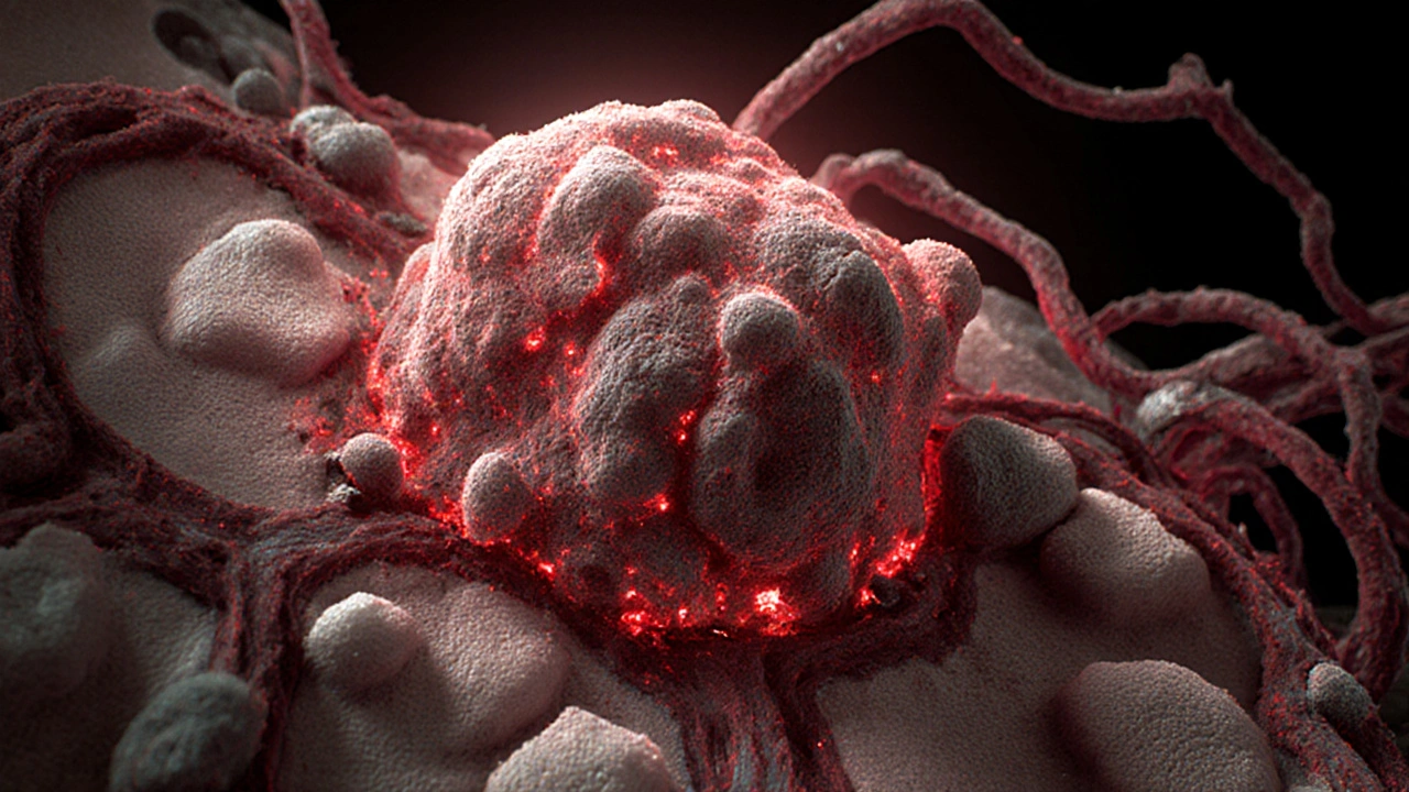
What is Cancer and How Does Tumor Growth Begin?
When doctors mention Cancer is a group of diseases marked by uncontrolled cell division, they’re referring to a cascade that starts at the cellular level. Normal cells obey signals that tell them when to grow, divide, or die. A mutation in the DNA can flip those signals, allowing a cell to ignore growth‑inhibiting cues. This faulty cell begins to multiply, forming a tumor a mass of abnormal cells that can be benign or malignant.
The initial phase of tumor growth is usually silent. Cells expand locally, forming a small nodule that may be undetectable on imaging. At this point, the lesion is often classified as benign because it lacks the ability to infiltrate surrounding tissue or spread to distant organs. The transition to malignancy hinges on several hallmarks: sustained proliferative signaling, evasion of growth suppressors, resistance to cell death, and the acquisition of replicative immortality.
Key Biological Drivers of Tumor Progression
Three processes dominate the shift from a localized tumor to an aggressive cancer:
- Carcinogenesis the transformation of normal cells into cancer cells through genetic and epigenetic changes - often spurred by carcinogens like tobacco smoke or UV radiation.
- Angiogenesis the formation of new blood vessels that feed the growing tumor - tumors release VEGF (vascular endothelial growth factor) to coax nearby vessels to sprout.
- Tumor Microenvironment the surrounding stromal cells, immune cells, and extracellular matrix that either hinder or support tumor growth - a supportive microenvironment can suppress immune attacks and promote invasion.
When these mechanisms align, the tumor gains the capacity to breach basement membranes, invade adjacent tissue, and eventually enter the bloodstream.
How Doctors Stage Cancer: The TNM System
Staging translates biological behavior into a practical language for treatment planning. The most widely used framework is the TNM Staging a classification that assesses Tumor size (T), lymph Node involvement (N), and Metastasis (M). Each component receives a numeric or alphabetical value, and the combination produces an overall stage from I (early) to IV (advanced).
| Component | Value | What It Means |
|---|---|---|
| T (Tumor) | T1‑T4 | Size and depth of the primary tumor |
| N (Nodes) | N0‑N3 | Number and location of regional lymph nodes involved |
| M (Metastasis) | M0 or M1 | Whether cancer has spread to distant organs |
Based on the TNM values, clinicians assign a stage:
- StageI: Small tumor (T1), no nodes (N0), no metastasis (M0).
- StageII: Larger tumor (T2‑T3) or minimal node involvement (N1) but still M0.
- StageIII: Significant node involvement (N2‑N3) or a tumor invading nearby structures; still no distant spread.
- StageIV: Any T, any N, but M1 - cancer has traveled to distant organs.
Characteristics of Each Cancer Stage
| Stage | Typical Tumor Size | Lymph Node Status | Metastasis | Usual Treatment | 5‑Year Survival (approx.) |
|---|---|---|---|---|---|
| I | ≤2cm | N0 | M0 | Surgery ± adjuvant therapy | 80‑90% |
| II | 2‑5cm | N0‑N1 | M0 | Surgery + chemotherapy/radiation | 60‑80% |
| III | >5cm or invading adjacent structures | N2‑N3 | M0 | Multimodal: surgery, chemo, radiation | 30‑60% |
| IV | Any | Any | M1 | Systemic therapy, targeted agents, palliative care | ≤10% |
These numbers are averages; individual outcomes vary based on cancer type, patient age, genetics, and response to therapy.

Why Metastasis Is the Game‑Changer
When cancer cells break away from the primary mass, they travel via blood or lymphatic vessels to seed new sites. This process, called Metastasis the spread of malignant cells to distant organs, forming secondary tumors, transforms a localized disease into a systemic one. The biology of metastasis involves epithelial‑to‑mesenchymal transition (EMT), loss of adhesion molecules like E‑cadherin, and the ability to survive in foreign tissue environments.
Clinically, the appearance of metastases pushes the disease to stageIV regardless of the original tumor size. It also signals the need for systemic treatments-chemotherapy, immunotherapy, or targeted agents-because local interventions (surgery, radiation) can’t remove cells dispersed throughout the body.
Linking Tumor Growth to Early Detection
Because tumor growth follows a predictable timeline-initiation, promotion, progression-screening programs aim to catch cancers in stageI or II. Imaging tools (mammography, low‑dose CT, MRI) and biomarkers (PSA, CA‑125, liquid biopsy cfDNA) provide snapshots of tumor size and metabolic activity. A small, non‑metastatic tumor is often curable with surgery alone, dramatically improving survival odds.
For example, breast cancer detection via annual mammography reduces mortality by about 30% in women aged 50‑69, primarily by finding tumors before they exceed 2cm (stageI). Similarly, low‑dose CT for high‑risk smokers catches lung nodules at a median size of 1.5cm, shifting many cases from stageIII to stageI.
Treatment Strategies Across Stages
Therapy is tailored to the stage because the goal evolves from eradication to control:
- StageI: Surgical excision with clear margins; sometimes radiation to reduce local recurrence.
- StageII: Surgery plus adjuvant chemotherapy; targeted therapy if molecular markers (e.g., HER2) are present.
- StageIII: Neoadjuvant chemotherapy to shrink tumors, followed by extensive surgery and postoperative radiation.
- StageIV: Systemic therapy (chemotherapy, immunotherapy, hormonal agents) aimed at slowing progression; palliative radiation to relieve symptoms.
Emerging approaches-CAR‑T cells, checkpoint inhibitors, and personalized vaccines-focus on the tumor microenvironment and aim to boost the immune system’s ability to recognize and destroy malignant cells, regardless of stage.
Common Pitfalls When Interpreting Staging Information
Even seasoned clinicians can stumble over staging nuances:
- Assuming size equals severity. A small tumor (T1) can already be N2 if lymph nodes are heavily involved.
- Mixing grade with stage. Grade describes how abnormal the cells look under a microscope, while stage reflects spread. High‑grade tumors often present at later stages, but not always.
- Overlooking molecular subtypes. Two stageII breast cancers may differ dramatically in prognosis based on hormone‑receptor status.
- Neglecting the patient’s overall health. Comorbidities can limit the feasibility of aggressive surgery, shifting the treatment plan despite a lower stage.
Being aware of these traps helps patients and families ask the right questions during consultations.
Next Steps for Patients and Caregivers
If you or a loved one has just received a cancer diagnosis, here’s a practical roadmap:
- Clarify the stage. Ask your oncologist to explain the TNM values and what they mean for prognosis.
- Request a pathology report. Look for tumor grade, hormone‑receptor status, and any genetic mutations (e.g., KRAS, EGFR).
- Discuss treatment options. Weigh surgery, systemic therapy, and clinical trial eligibility based on stage and molecular profile.
- Consider a second opinion. A fresh set of eyes can confirm staging accuracy and suggest alternative approaches.
- Plan supportive care. Nutrition, physical therapy, and mental‑health resources improve tolerance to treatment and quality of life.
Staying informed about how tumor growth translates into staging empowers you to make decisions that align with your goals and values.
Frequently Asked Questions
What distinguishes a benign tumor from a malignant one?
Benign tumors grow slowly, stay confined, and rarely invade nearby tissue or spread. Malignant tumors invade, destroy surrounding structures, and can metastasize through blood or lymph vessels.
How is the TNM system applied to different cancer types?
Each organ has specific T‑categories (e.g., T1 for a breast tumor ≤2cm, T1 for a lung tumor ≤3cm). N and M definitions also vary, but the underlying principle-size, nodal involvement, distant spread-remains consistent across cancers.
Can cancer be staged without imaging?
Imaging (CT, MRI, PET) provides crucial data on tumor size and metastasis. In some early‑stage cases, physical exam and pathology may suffice, but imaging is generally required for accurate staging.
What role do genetic mutations play in staging?
Genetic alterations don’t change the anatomical stage, but they affect treatment choices and prognosis. For instance, an EGFR mutation in lung cancer may make a patient eligible for targeted therapy, improving outcomes even in stageIII.
Is stageIV always incurable?
StageIV denotes metastatic disease, which is generally not curable with current technology. However, many patients achieve long‑term control and meaningful quality of life using modern systemic therapies.
Jordan Schwartz
September 29, 2025 AT 13:28Understanding how the TNM system breaks down tumor size, nodal involvement, and metastasis really helps patients see a clear roadmap for treatment decisions.
When you know a tumor is T1 N0 M0, the outlook is often very optimistic with surgery alone.
Even a stage III diagnosis can be tackled with multimodal therapy, giving many hope for extended survival.
Keep focusing on early detection; it makes a world of difference.
Nitin Chauhan
September 29, 2025 AT 19:01Knowing the stage means you can plan the right treatment path. Tumor size matters but nodes tell the story too. Metastasis is the game changer and flips everything. Early screening catches cancers before they hit M1. Stay proactive and get checked regularly.
Angelo Truglio
September 30, 2025 AT 00:35Look! The medical community has dressed up a simple truth in layers of jargon!!! They parade the TNM system as if it were a flawless oracle, yet we see countless lives tangled in its bureaucracy!!! Ignorance of real risk factors is a sin we cannot overlook; patients deserve crystal‑clear communication, not cryptic codes!!!
Dawn Midnight
September 30, 2025 AT 06:08The staging criteria are defined by strict anatomical parameters; any deviation from these measurements must be documented meticulously. Accurate assessment of T, N, and M values ensures consistent treatment planning across institutions. It is essential to differentiate between size and biological aggressiveness when interpreting stage.
frank hofman
September 30, 2025 AT 11:41lol i guess the whole staging thing is just a way to scare u 😜 but honestly some ppl get stage IV and live longer than stage II folks 😂 it’s all about the vibe not the numbers
Dannii Willis
September 30, 2025 AT 17:15While the TNM framework provides a solid baseline, integrating patient preferences can personalize care pathways. It’s useful to discuss both the statistical outcomes and the individual’s lifestyle goals when reviewing stage‑specific options.
Robyn Du Plooy
September 30, 2025 AT 22:48From an oncological systems biology perspective, the interplay between angiogenic signaling pathways and the tumor microenvironment modulates the transition from T2 to T3 lesions. Quantitative imaging biomarkers, such as perfusion MRI, can refine nodal staging beyond the conventional N1/N2 dichotomy.
Boyd Mardis
October 1, 2025 AT 04:21Stage III is the battlefield where every treatment arm fights for dominance.
ayan majumdar
October 1, 2025 AT 09:55yeah early check ups save lives they catch stuff before it spreads
Sarah Aderholdt
October 1, 2025 AT 15:28Understanding the philosophical implications of labeling a disease by stage reshapes how we perceive mortality.
Phoebe Chico
October 1, 2025 AT 21:01Our nation’s strength lies in empowering patients with knowledge; the TNM system is a tool, not a tyrant. Embrace the science, reject the fatalism, and champion accessible screening for every citizen.
Larry Douglas
October 2, 2025 AT 02:35The TNM classification, established by the Union for International Cancer Control, utilizes a hierarchical schema where T denotes primary tumour dimensions, N represents regional lymph node involvement, and M indicates distant metastatic spread. This trichotomous model underpins most clinical trial eligibility criteria and informs prognostic modeling.
Ann Campanella
October 2, 2025 AT 08:08Stage IV is still fightable.
Desiree Tan
October 2, 2025 AT 13:41Don’t let a diagnosis define your limits; use it as a catalyst for proactive health management. Harness the power of multidisciplinary teams to attack the disease from every angle. Set concrete goals for each treatment phase and celebrate every milestone, no matter how small. Your determination fuels the therapeutic arsenal.
Andrea Dunn
October 2, 2025 AT 19:15They don’t tell you that the staging data is often manipulated by pharma giants to push expensive drugs 😒 keep your eyes open and question every statistic.
Erin Johnson
October 3, 2025 AT 00:48It’s fascinating how the layperson’s panic over a “stage IV” label often eclipses the nuanced reality of modern oncology. While many assume that metastasis seals an inevitable fate, recent advances in immunotherapy have turned some advanced cancers into chronic, manageable conditions. The TNM system, though imperfect, provides a common language that bridges research, clinical trials, and bedside decisions. Yet critics love to harp on its inability to capture molecular heterogeneity, a point that, frankly, is hardly shocking given the system’s anatomical roots. In practice, clinicians combine stage information with genomic profiling to tailor targeted agents, offering a glimmer of hope that stage alone cannot convey. Moreover, early detection programs have demonstrably shifted populations toward earlier stages, boosting five‑year survival rates dramatically. Consider breast cancer: routine mammography has nudged a substantial fraction of cases into stage I, where surgery alone often suffices. On the other hand, lung cancer screening with low‑dose CT has similarly up‑staged diagnoses from late to early, saving countless lives. The key takeaway is that stage is a stepping stone, not a death sentence. Patients should be encouraged to seek second opinions, explore clinical trial options, and stay informed about emerging therapies. And yes, while the word “metastasis” does sound ominous, it merely flags the need for systemic treatment rather than doom. So, keep the sarcasm handy when friends dramatize cancer stages, but also respect the genuine anxiety that a new diagnosis can provoke. Knowledge empowers, and humor, when used wisely, can lighten the heavy load. Ultimately, a balanced perspective-rooted in scientific evidence and compassionate support-makes the journey more bearable. Stay proactive, stay hopeful, and let your care team guide you through each phase.
Rica J
October 3, 2025 AT 06:21Hey there, just wanted to say the staging thing can sound super confusing at first but once you get the hang of T N and M it gets easier.
Make sure you ask your doc about any weird terms they use – better ask than guess.
And don’t forget, staying on top of your appointments can really make a difference.
Linda Stephenson
October 3, 2025 AT 11:55Ever wonder how different cancers use the same TNM language yet behave so uniquely? Sharing resources and personal stories can help everyone navigate the maze together.
Sunthar Sinnathamby
October 3, 2025 AT 17:28Take charge of your health journey by learning the stage details and demanding the best care options! Push for early screening, stay active, and rally your support network – the fight is yours to win.