CT Angiography – Overview, Uses, and Risks
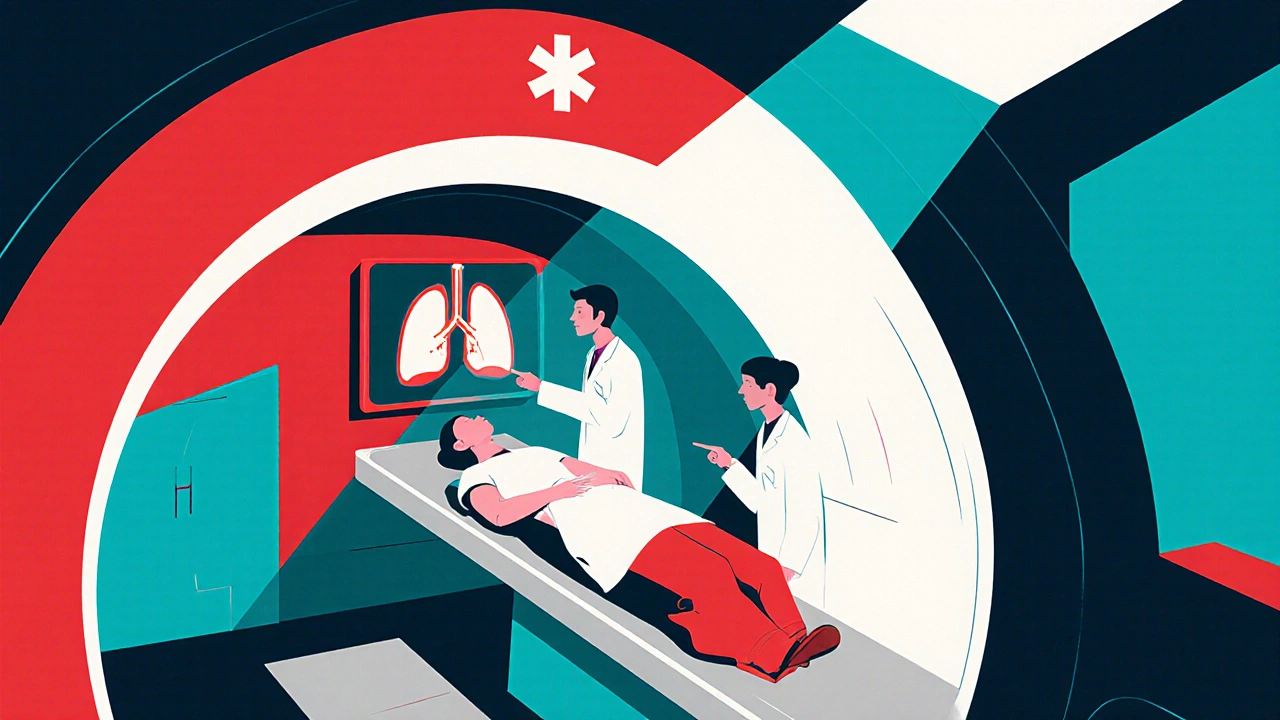
When you hear CT angiography, a medical imaging technique that combines computed tomography scanning with injected contrast material to visualize blood vessels in high detail. Also known as CTA, it lets doctors see arteries and veins without an invasive procedure. The method builds on computed tomography, a cross‑sectional X‑ray technology that creates detailed 3‑D images of the body, and pairs it with contrast media, a iodine‑based dye that highlights vascular structures during the scan. Together, they enable precise vascular imaging, the visual assessment of blood flow and vessel integrity, which is essential for diagnosing conditions like coronary artery disease, aortic aneurysms, and pulmonary embolism.
Why do doctors reach for CT angiography over other tests? First, it offers speed – a full‑body vessel map can be produced in under a minute, making it ideal for emergency settings such as suspected pulmonary embolism where time is critical. Second, the image resolution is high enough to spot tiny blockages that might be missed on standard CT scans, so it’s a go‑to tool for planning heart‑related interventions like stent placement. Third, because the scan captures the whole vascular tree in one session, it reduces the need for multiple appointments, saving both time and cost. In practice, the workflow looks like this: a patient receives an injection of contrast media, the CT scanner rotates around them, and sophisticated software reconstructs the data into 3‑D views that radiologists interpret. This chain—contrast injection, CT acquisition, and 3‑D reconstruction—creates a clear semantic triple: CT angiography combines CT scanning with contrast media to produce vascular imaging.
Key Benefits and Practical Considerations
While the diagnostic power of CT angiography is impressive, it’s not without trade‑offs. The main concern is radiation exposure; a typical CTA delivers a dose roughly equivalent to a few months of natural background radiation. Modern scanners, however, use dose‑reduction algorithms that cut exposure by up to 50 % without compromising image quality. Another factor is the use of iodine‑based contrast, which can cause allergic reactions or kidney stress in vulnerable patients. Before the scan, clinicians usually check kidney function and ask about prior contrast allergies. For those at risk, alternatives like magnetic resonance angiography (MRA) may be offered, though MRA takes longer and isn’t as widely available in emergency rooms. Finally, patient preparation matters: staying still, holding breath at the right moment, and following any fasting instructions help ensure clear images and reduce the chance of repeat scans.
The breadth of topics covered by our collection reflects how CT angiography fits into everyday clinical decisions. Below you’ll find articles that dive into specific drug interactions that matter when contrast media is used, comparisons of blood‑pressure medications that affect vascular health, and guides on managing side‑effects of treatments for lung‑related diseases— all relevant for patients who might undergo a CTA. Whether you’re a clinician looking for quick reference points or a patient preparing for the test, the posts that follow give practical insights, safety tips, and up‑to‑date evidence to help you navigate the whole process confidently.
CT Scans in Embolism Diagnosis and Management: What You Need to Know
Explore how CT scans detect and guide treatment of embolisms, from pulmonary clots to arterial blockages, with practical tips, pitfalls, and a CTA vs V/Q comparison.
Read more